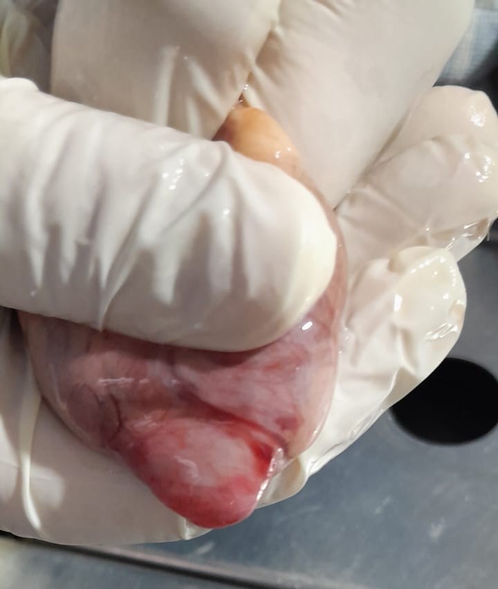
Apical aneurysm of the feline left ventricle. Literature review based on a series of 17 cases
Apical aneurysm of the feline left ventricle. Literature review based on a series of 17 cases
DOI:
https://doi.org/10.19137/cienvet.v28.9866Keywords:
Feline cardiomyopathy, mid-ventricular hypertrophy, AneurysmAbstract
Left mid-ventricular obstruction (LMVO) is a sparsely documented entity in veterinary medicine. In human medicine, it is associated with the development of apical aneurysms (AA) due to flow entrapment and high apical systolic pressure. The objective of this study is to describe the correlation between echocardiographic and anatomopathological findings in a series of 17 felines with LMVO and AA. Seventeen felines (aged 8–15 years; 55% male and 45% female) diagnosed with LMVO and AA between 2015 and 2024 were evaluated. Fourteen patients were diagnosed pre-mortem via Doppler echocardiography, and three post-mortem. Seven necropsies were performed with histopathological studies (H&E and Masson's Trichrome) of the ventricular wall and the left anterior descending artery. LMVO was caused by myocardial hypertrophy, fibromuscular bands/membranes, or excess false tendons. All patients were normotensive and presented no outflow tract obstruction. Color Doppler detected mid-ventricular turbulent flow in 100% of the live cases. Histopathology revealed fibrosis in the aneurysm wall without evidence of obstructive coronary disease, suggesting a hemodynamic etiology. AAs associated with hypertrophic cardiomyopathy showed greater size and dense fibrosis compared to other causes of obstruction. The association between LMVO and the development of AA in felines is confirmed. The absence of coronary lesions reinforces the theory of flow entrapment as a mechanical cause of myocardial injury. This represents the largest case series reported to date, constituting a significant contribution to feline and comparative cardiology
References
1.MacLea H, Boon J, Bright J. Doppler Echocardiographic Evaluation of Midventricular Obstruction in Cats with Hypertrophic Cardiomyopathy. J. Vet. Intern; 2013, 27(6), 1416-1420. https://doi.org/10.1111/jvim.12175
2.Forero S,Moreno NL. Miocardiopatía hipertrófica septal, la gran simuladora. APCyCCV,2021: 274-278. https://doi.org/10.47487/apcyccv.v2i4.1
3. Fox PR. Hypertrophic cardiomyopathy. Clinical and pathologic correlates. J Vet Cardiol. 2003 nov;5(2):39-45. doi:10.1016/S1760-2734(06)70051-0
4. Alcaino I. Milton, Aguilar P. Juan, Jorquera B. Claudia. Aneurisma apical en paciente con miocardiopatía hipertrófica. Rev. méd. Chile [Internet]. 2021 Mar [citado 2026 Jun 29]; 149(3): 472-475. Disponible en: http://www.scielo.cl/scielo.php?script=sci_arttext&pid=S0034-98872021000300472&lng=es. http://dx.doi.org/10.4067/s0034-98872021000300472.
5. Acquaviva A, et al. ACVIM consensus statement: Guidelines for the identification, evaluation, and management of systemic hypertension in dogs and cats . J Vet Intern Med. 2018;32(6):1815-1833.https://pmc.ncbi.nlm.nih.gov/articles/PMC6271319/
6. Deng L, Zuo H, Li A, Yang C, Huang X. Numerical Simulation Study on the Mechanism of Formation of Apical Aneurysm in Hypertrophic Cardiomyopathy With Midventricular Obstruction. Front Physiol. 2021 jul 21; 12:717717. doi: 10.3389/fphys.2021.717717. PMID: 34366902; PMCID: PMC8334850.
7. Strachinaru M, Huurman R, Bowen DJ, Schinkel AFL, Hirsch A, Michels M. Relation Between Early Diastolic Mid-Ventricular Flow and Elastic Forces Indicating Aneurysm Formation in Hypertrophic Cardiomyopathy. J Am Soc Echocardiogr. 2022 Aug;35(8):846-856.e2. doi: 10.1016/j.echo.2022.04.010. Epub 2022 Apr 27. PMID: 35489541.
8. Altay H, Altin C, Coner A, Muderrisoglu H. Normal coronary artery patient presenting with left ventricular aneurysm. Case Rep Med. 2011; 2011:183050. doi: 10.1155/2011/183050. Epub 2011 Aug 7. PMID: 21845194; PMCID: PMC3154388.
9. Cianciulli TF et al. Subaortic and mid-ventricular obstructive hypertrophic cardiomyopathy with an apical aneurysm: a case report. Cardiovasc Ultrasound. 2006 Mar 22; 4:15. doi: 10.1186/1476-7120-4-15. PMID: 16553961; PMCID: PMC1501052.
10. Efthimiadis GK et al. Clinical characteristics and natural history of hypertrophic cardiomyopathy with midventricular obstruction. Circ J. 2013;77(9):2366-74. doi: 10.1253/circj. cj-12-1561. Epub 2013 May 31. PMID: 23728066.
11. Lee DZJ et al. Clinical Characteristics and Prognostic Importance of Left Ventricular Apical Aneurysms in Hypertrophic Cardiomyopathy. JACC Cardiovasc Imaging. 2022 oct;15(10):1696-1711. doi: 10.1016/j.jcmg.2022.03.029. Epub 2022 Jun 15. PMID: 36202449.
12. Maron MS et al. Prevalence, clinical significance, and natural history of left ventricular apical aneurysms in hypertrophic cardiomyopathy. Circulation. 2008 oct 7;118(15):1541-9. Epub 2008 Sep 22. PMID: 18809796. Doi: 10.1161/CIRCULATIONAHA.108.781401
13. Rowin EJ et al. Hypertrophic Cardiomyopathy With Left Ventricular Apical Aneurysm: Implications for Risk Stratification and Management. J Am Coll Cardiol. 2017 Feb 21;69(7):761-773. doi: 10.1016/j.jacc.2016.11.063. Erratum in: J Am Coll Cardiol. 2017 Mar 28;69(12):1652. doi: 10.1016/j.jacc.2017.02.014. PMID: 28209216.
14. Urbano-Moral JA, Lopez-Haldon JE. Aspectos poco conocidos de la miocardiopatía hipertrófica. Revista Española de Cardiología, (2015) 68(7), 554-558. https://doi.org/10.1016/j.recesp.2015.03.010
15. Sherrid MV et al. Apical Aneurysms and Mid-Left Ventricular Obstruction in Hypertrophic Cardiomyopathy. JACC Cardiovasc Imaging. 2023 May;16(5):591-605. doi: 10.1016/j.jcmg.2022.11.013. Epub 2023 Jan 11. PMID: 36681586.
16. Strachinaru M et al. Relation Between Early Diastolic Mid-Ventricular Flow and Elastic Forces Indicating Aneurysm Formation in Hypertrophic Cardiomyopathy. J Am Soc Echocardiogr. 2022 Aug;35(8):846-856.e2. doi: 10.1016/j.echo.2022.04.010.
17. Tomić S et al. Characteristics of Akinetic and Dyskinetic Left Ventricular Aneurysms in the Context of Echocardiographic Diagnosis and Treatment Selection. Medicina (Kaunas). 2024 jul 16;60(7):1141. doi: 10.3390/medicina60071141.
18. Tomić S et al. Comparative Analysis of Basal vs. Apical Left Ventricular Aneurysms: Impact on Ejection Fraction and Cardiac Function. Medicina (Kaunas). 2024 Sep 26;60(10):1578. doi: 10.3390/medicina60101578.
19. Fan HG, Zheng Z, Feng W, Yuan X, Wang W, Hu SS. Repair of left ventricular aneurysm: ten-year experience in Chinese patients. Chin Med J (Engl). 2009 Sep 5;122(17):1963-8. DOI: 10.3760/cma.j.issn.0366-6999.2009.17.001
20. Perillo EF et al. Diagnosis and Clinical Implication of Left Ventricular Aneurysm in Hypertrophic Cardiomyopathy. Diagnostics (Basel). 2023 May 25;13(11):1848. doi: 10.3390/diagnostics13111848.
21. Cesta MF, Baty CJ, Keene BW, Smoak IW, Malarkey DE. Pathology of end-stage remodeling in a family of cats with hypertrophic cardiomyopathy. Vet Pathol. 2005 jul;42(4):458-67. doi: 10.1354/vp.42-4-458.
22. Guarda F, Tursi M, Bravo M, Spaino G. Gli aneurismi cardiaci ventricolari negli animali. Vetjournal.it, Large Animals Review, Anno 10, n. 2, Aprile 2004.
23. Herráez P, Rodríguez F, Aguirre-Sanceledonio M, Edwards JF, Suárez-Bonnet A, Espinosa de Los Monteros A. Congenital biventricular cardiac diverticula in a dog. Vet Pathol. 2011. https://doi.org/10.1177/0300985810375243
24. Novo Matos J et al. Thin and hypokinetic myocardial segments in cats with cardiomyopathy. J Vet Cardiol. 2023 Apr; 46:5-17. doi: 10.1016/j.jvc.2023.02.002.
25. Toschi Corneliani R, Tursi M, Spalla I. Advanced imaging findings in a cat with left ventricular apical aneurysm. J Vet Cardiol. 2022 oct; 43:55-60. doi: 10.1016/j.jvc.2022.06.008.
26. Williams KJ. Coronary arteriosclerosis with myocardial atrophy in a 13-year-old dog. Vet Pathol. 2003 nov;40(6):695-7. doi: 10.1354/vp.40-6-695.
Published
Issue
Section
License
Copyright (c) 2026 Guillermo Claudio Belerenián, Pablo Alejandro Donati, Victor Castillo, Cristian Daniel Rodriguez, Gustavo Abuin, Juan Manuel Guevara

This work is licensed under a Creative Commons Attribution-NonCommercial-ShareAlike 4.0 International License.
Al momento de enviar sus contribuciones, los colaboradores deberán declarar , de manera fehaciente, que poseen el permiso del archivo o repositorio donde se obtuvieron los documentos que se anexan al trabajo, cualquiera sea su formato (manuscritos inéditos, imágenes, archivos audiovisuales, etc.), permiso que los autoriza a publicarlos y reproducirlos, liberando a la revista y sus editores de toda responsabilidad o reclamo de terceros , los autores deben adherir a la licencia Creative Commons denominada “Atribución - No Comercial CC BY-NC-SA”, mediante la cual el autor permite copiar, reproducir, distribuir, comunicar públicamente la obra y generar obras derivadas, siempre y cuando se cite y reconozca al autor original. No se permite, sin embargo, utilizar la obra con fines comerciales.
How to Cite





.jpg)